|
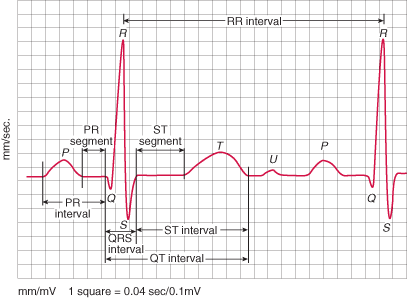
Ģ ItÆs a small wave that follows the T-wave when visible Ģ Not all rhythm strips will show a U-wave. Ģ Generally we donÆt measure or chart on a U-wave. Ģ They mostly become visible when the heart rate falls below 60. Ģ ItÆs a little mysterious in that its origin or source is unknown. |
|
In general, itÆs best to know what a U-wave is, and that itÆs just there tagging along following the T-wave. Typically, when a Sinus Rhythm slows down below 60, (Sinus Brady) the U-wave becomes more prominent. Sinus Brady with a strong U-wave can sometimes fool you into thinking you have a dropped beat (2nd degree AV block). If you arenÆt sure, see that the P-waves march out or not. P-waves should march out for 2nd degree block, and normal U-waves typically have a wider lower shape to them. For diagnosing or determining treatment of any kind, that will be up to Cardiology. This short study is for simply knowing what it is. For further information, we have included much more on abnormal U-waves listed down below. Ģ Did you know that with patients presenting chest pain, inverted U-waves are a very specific sign of Myocardial Ischemia Ģ The Same conditions that cause a prominent (large) U-wave will also cause a long QT segment. |
|
Inverted U-waves Ģ U-wave inversion is abnormal (in leads with upright T waves) Ģ A negative U wave is highly specific for the presence of heart disease The main causes of inverted U waves are: Ģ Coronary artery disease Ģ Hypertension Ģ Valvular heart disease Ģ Congenital heart disease Ģ Cardiomyopathy Ģ Hyperthyroidism The source of the U wave is unknown. Theories regarding its origin are: Ģ Delayed repolarisation of Purkinje fibres Ģ Prolonged repolarisation of mid-myocardial ōM-cellsö Ģ After-potentials resulting from mechanical forces in the ventricular wall Features of Normal U-waves Ģ The U wave normally goes in the same direction as the T wave Ģ U -wave size is inversely proportional to heart rate: the U wave grows bigger as the heart rate slows down Ģ U waves generally become visible when the heart rate falls below 65 bpm Ģ The voltage of the U wave is normally less than 25% of the T-wave voltage: disproportionally large U waves are abnormal Ģ Maximum normal amplitude of the U wave is 1-2 mm. Abnormalities of the U-wave Ģ Prominent U waves Ģ Inverted U waves Prominent U-waves Ģ U waves are prominent if > 1-2mm or 25% of the height of the T wave. Ģ The most common cause of prominent U waves is bradycardia. Ģ Abnormally prominent U waves are characteristically seen in severe hypokalaemia. Prominent U-waves may also be seen with: Ģ Hypocalcaemia Ģ Hypomagnesaemia Ģ Hypothermia Ģ Raised intracranial pressure Ģ Left ventricular hypertrophy Ģ Hypertrophic cardiomyopathy The following drugs may cause prominent U-waves: Ģ Digoxin Ģ Phenothiazines (thioridazine) Ģ Class Ia antiarrhythmics (quinidine, procainamide) Ģ Class III antiarrhythmics (sotalol, amiodarone) |
Return to Home Page
Refrences and graphics were used from: http://lifeinthefastlane.com/ecg-library/basics/u-wave/
I wave, You wave, we all wave for U-waves ¢ (yes goofy indeed)
